Normal CT Head
Interactive normal CT head anatomy viewer with scrollable axial slices. Identify ventricles, basal ganglia, and skull base landmarks on non-contrast CT.
Explore this normal CT head to get up to speed on anatomy.
How to use the viewer
The viewer has three modes, accessible via the tabs in the right panel: Explore, Quiz, and Report.
Explore mode. Scroll through all 41 axial slices using your mouse wheel, the slider at the bottom, or the arrow keys (J/K). Over 50 labeled structures are listed in the panel — click any name to highlight it on the image with a color overlay. This is the best way to orient yourself to unfamiliar anatomy: pick a structure, highlight it, then scroll up and down to see how its appearance changes across slices.
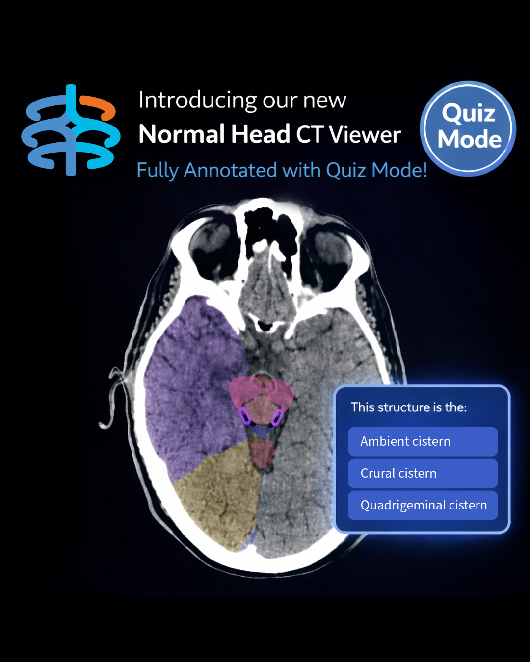
Quiz mode. Test yourself with multiple-choice questions that highlight a structure on the image and ask you to identify it. Get five correct answers in a row to earn a streak. Hit the spacebar to advance to the next question without reaching for the mouse — this makes rapid-fire drilling efficient.
Report mode. Read a structured radiology report for this study, organized the way a radiologist would dictate it: clinical context, then findings broken out by brain, CSF, sinuses, bones, orbits, and soft tissue, followed by an impression. This gives you a model for what a normal report looks like and how findings are organized by anatomic region.
What you're looking at
CT images of the brain are displayed in the axial plane and viewed from below, as if you were standing at the patient's feet and looking up. The right side of the brain appears on the left side of the screen. Anterior structures are at the top of the image.
On a non-contrast study, tissue density determines brightness. Bone and acute blood appear bright white. Gray matter is slightly brighter than white matter. Cerebrospinal fluid (CSF) in the ventricles and cisterns appears dark. Air in the paranasal sinuses and mastoid air cells is black.
Key anatomy by level
Skull base and posterior fossa. The lowest slices show the posterior fossa structures: the cerebellar hemispheres and vermis, the brainstem (medulla, pons, and midbrain), and the cerebral peduncles. The fourth ventricle sits between the cerebellum and brainstem, with the cerebral aqueduct of Sylvius connecting it superiorly to the third ventricle. Surrounding CSF spaces include the interpeduncular cistern and quadrigeminal cistern. The cervicomedullary junction and cervical spinal cord are visible on the lowest slices. Bony structures at this level include the mastoid air cells, the sphenoid sinuses, and the petrous bones. The nasopharynx, oropharynx, and oral cavity are also seen anteriorly.
Orbits and face. Slightly higher, the globes and lenses come into view within the orbits, along with the optic nerves coursing posteriorly toward the chiasm. The nasal cavity, nasal septum, ethmoid sinuses, maxillary sinuses, and frontal sinuses are all visible at their respective levels. The infratemporal fossa is seen laterally.
Suprasellar region and basal ganglia. Moving superiorly, the suprasellar cistern appears as a star-shaped dark CSF space. The basal ganglia come into view: the caudate head, putamen, and globus pallidus are gray matter nuclei flanking the internal capsule, which is divided into an anterior limb, genu, and posterior limb. The thalamus sits medially, separated from its counterpart by the slit-like third ventricle. The insular cortex is visible deep within the Sylvian fissures. The middle cerebral arteries (MCA) and basilar artery can be identified at this level. The ambient cisterns and crural cisterns wrap around the midbrain.
Ventricles. The ventricles are a series of interconnected CSF-filled chambers within the brain. The two lateral ventricles are the largest, each with a frontal horn, body, atrium, occipital horn, and temporal horn. They connect to the midline third ventricle through the paired foramina of Monro. The third ventricle, a narrow slit between the thalami, communicates with the fourth ventricle via the cerebral aqueduct of Sylvius. The fourth ventricle sits in the posterior fossa between the cerebellum and brainstem. CSF exits the fourth ventricle laterally through the foramina of Luschka into the premedullary cistern, inferiorly through the foramen of Magendie into the cisterna magna, and caudally through the obex into the central canal of the spinal cord. Obstruction at any of these connection points can cause hydrocephalus — a high-yield finding to recognize on CT. The choroid plexus, which produces CSF, is often visible as calcified bright foci within the atria of the lateral ventricles.
Cerebral hemispheres and vertex. At the level of the lateral ventricle bodies, the corpus callosum connects the hemispheres — the genu anteriorly and the splenium posteriorly. The frontal, temporal, parietal, and occipital lobes are all distinguishable. Higher still, the precentral and postcentral gyri straddle the central sulcus — a key landmark for the primary motor and sensory cortices. The highest slices show the cerebral convexities with their gyral-sulcal pattern. Gray-white differentiation should be clearly visible, with cortical gray matter slightly brighter than the underlying white matter.
Dural venous sinuses. The superior sagittal sinus runs along the midline at the top of the falx. The transverse sinuses course laterally along the posterior fossa, and the sigmoid sinuses descend toward the jugular foramina. The torcula (confluence of sinuses) is the junction point where these major sinuses converge at the internal occipital protuberance. The vertebral arteries are visible at the skull base.
Common pitfalls when learning CT anatomy
Confusing gray and white matter. On CT, gray matter is slightly brighter than white matter — the opposite of their names. The cortical ribbon (gray matter) lines the surface, while the deeper white matter tracts appear slightly darker. Losing track of this relationship is one of the most common early mistakes. Use Explore mode to click between cortical and subcortical structures and compare their densities.
Forgetting the skull base structures. Learners tend to focus on the cerebral hemispheres and skip the lowest slices. The posterior fossa contains critical anatomy — cerebellum, brainstem, fourth ventricle — and is where many life-threatening pathologies present. Make a habit of starting your review from the bottom.
Misidentifying the basal ganglia components. The caudate head, putamen, globus pallidus, and thalamus are all gray matter nuclei deep in the brain, but they sit in different locations relative to the internal capsule. Use the internal capsule as your dividing landmark: caudate and putamen are lateral, thalamus is medial and posterior. In Explore mode, clicking these structures individually and toggling between them is the fastest way to lock in their spatial relationships.
Overlooking the cisterns. The basal cisterns — ambient, crural, interpeduncular, and quadrigeminal — are often skipped by beginners. These CSF spaces are important landmarks and their effacement is an early sign of mass effect or herniation. The viewer labels each cistern individually so you can see exactly where they sit relative to the brainstem.
Not tracing the ventricles as a connected system. It is easy to identify the lateral ventricles in isolation but forget that they connect to the third ventricle (via the foramina of Monro), which connects to the fourth ventricle (via the aqueduct). Tracing this pathway on every study reinforces the anatomy and helps you spot early hydrocephalus.
Mixing up lobes and fissures. The Sylvian fissures separate the frontal and temporal lobes. The central sulcus separates the precentral gyrus (motor) from the postcentral gyrus (sensory). These landmarks are labeled in the viewer — practice identifying them before scrolling to confirm.
Why this matters for your clinical training
Whether you are a PA student, NP student, or medical student, being comfortable with normal CT head anatomy is foundational. On clinical rotations — especially in emergency medicine, neurology, and surgery — you will be expected to look at head CTs and identify obvious abnormalities. That starts with knowing what normal looks like.
Use this viewer to scroll through the study multiple times. On each pass, focus on a different system: trace the ventricles, follow the cortical sulci, identify the basal cisterns, or map the dural venous sinuses. Then switch to Quiz mode and test your retention. Repetition builds pattern recognition.