The case of radiating chest pain

Hello, everyone! In this week’s edition of "This Week in Radiology," we're focusing on a case that highlights the importance of knowing when to push for more imaging. Let's take a closer look at a 60-year-old hypertensive man who came into the ER with severe chest pain radiating to his upper back.
Getting Started
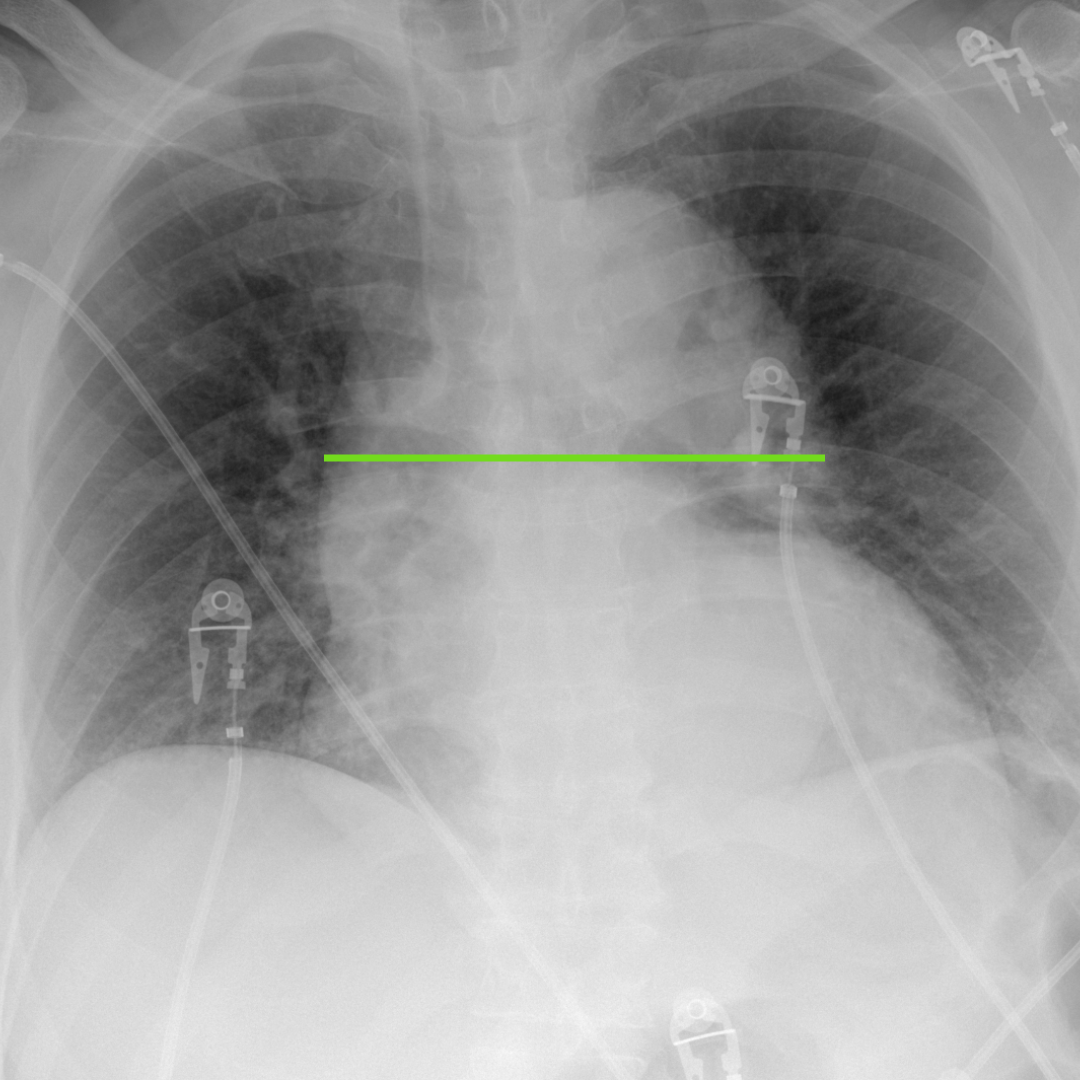
Our initial investigation began with a history and physical exam with a suspicion for a variety of acute conditions like heart attack, aortic dissection, and pneumothorax. We ordered the portable chest radiograph, because it's a fast and cost-effective screening tool whenever you suspect issues with the thorax. In this case, the chest radiograph showed an enlarged cardiomediastinal silhouette. Unfortunately, you'll see a mediastinum like this in the spectrum of normal patients, particularly with portable radiographs. While these signs were helpful, they didn't give us a complete picture. In radiology, it's often about piecing together different bits of information to get a clearer understanding.
Deeper Insight: CT Aortogram
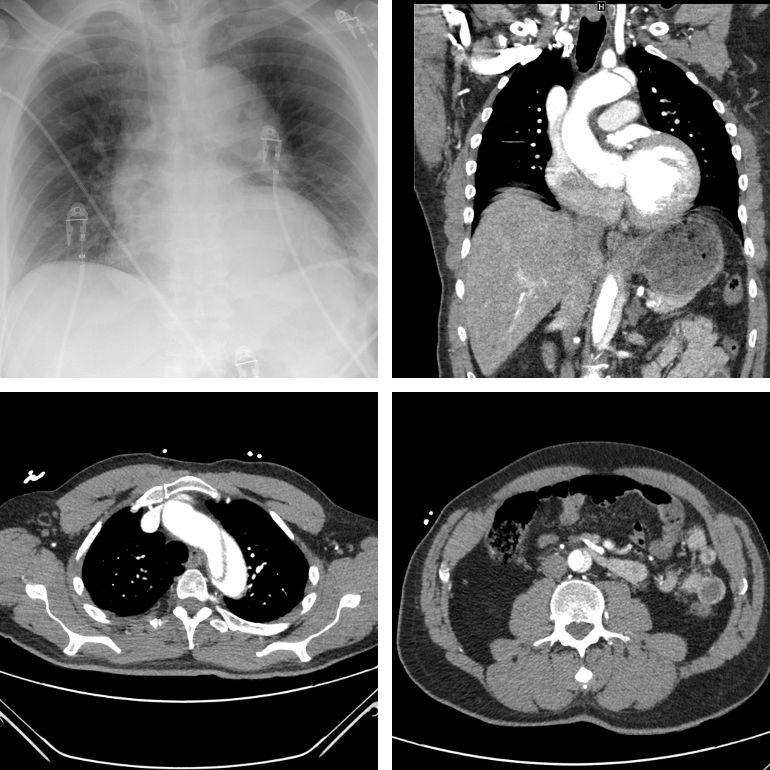
Given the nature of the patient's symptoms, we proceeded with a CT aortogram. Using intravenous iodinated contrast, the CT aortogram provided more detail on the aorta, and revealed an aortic dissection at the level of the aortic arch and abdominal aorta. This was clearly marked with an intimal flap, distinguishing the true and false lumens – a classic sign of this condition.
Educational Takeaway
For our future medical professionals, this case underscores a valuable point: in situations with a high clinical suspicion for conditions like aortic dissection, relying solely on a CXR may not be sufficient. The CTA plays a crucial role in such scenarios, offering the precision and detail needed for an accurate diagnosis.
Each week, we aim to bring you insights that not only broaden your knowledge but also prepare you for the practical realities of medical diagnostics. As always, stay engaged, keep learning, and we look forward to sharing more insights with you next week in "This Week in Radiology." Stay tuned! 📚👩⚕️👨⚕️🔬
"This Week in Radiology" is provided for educational purposes. Elements of the patient's history have been modified for privacy while retaining a realistic clinical presentation relative to the radiological findings. Any decision-making for specific patients on your service should be made by the patient's expert team, accounting for the specific context of that patient and practice environment.