The case of a suddenly painful chest
December 15, 2023
This week in radiology, we saw a young man present to the emergency room with sudden-onset chest pain after extensive coughing. As far as imaging goes, the chest radiograph is a good place to start, because it can catch related findings like pneumothorax, infiltrates, and peribronchial thickening. But this case is a reminder that you'll need to be sharp and a little bit lucky to catch more subtle signs of an equally important complication—pneumomediastinum.
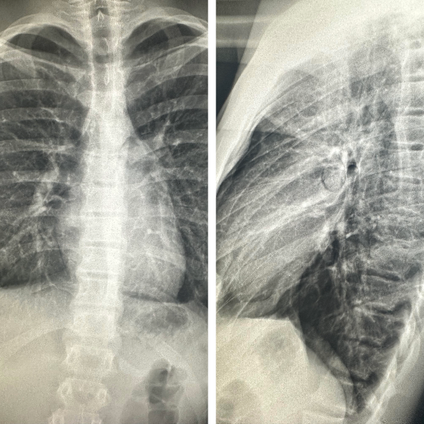
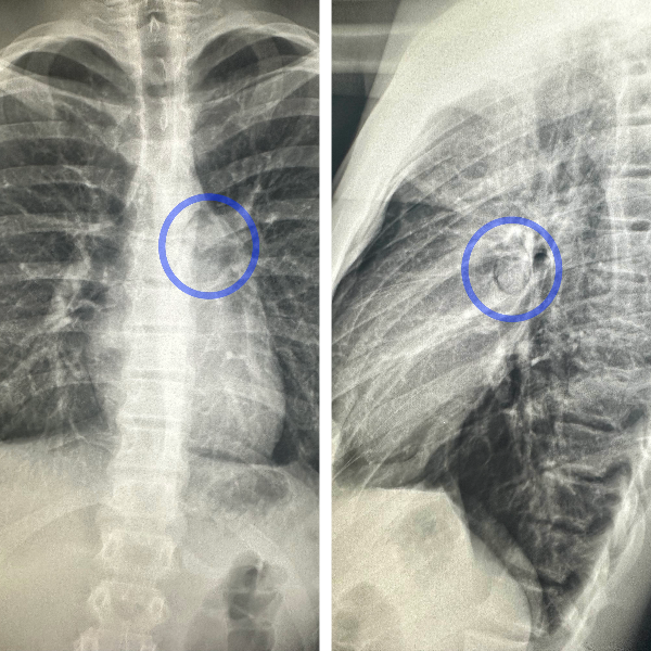
Take a look at the cardiomediastinum in the PA radiograph. The classic appearance for pneumomediastinum is a sliver of lucency along the left cardiac border or upper mediastinum. But there is no definite indication of that in this patient. Instead, the frontal radiograph gives us a very subtle lucency near the left hilum, that's probably too subtle to catch on its own. On the other hand, the lateral radiograph is the slam dunk. At the hilum, we saw a circular arc of lucency, enough of an abnormality to order a chest CT.
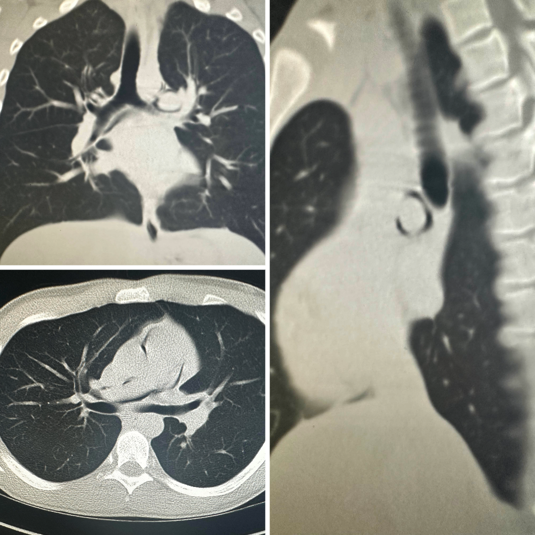
The chest CT confirmed our suspicion for pneumomediastinum. Here you see coronal and satittal cuts through the mediastinum, demonstrating the arcs of air are adjacent to the mainstem bronchi and pulmonary arteries.
There are many causes for pneumomediastinum, including spontaneous, barotrauma, esophageal perforation, and infection. Finding pneumomediastinum on a chest radiograph can be hard to do, but will also reduce the patient's time to diagnosis and hasten their way to a judicious diagnosis and treatment.
—RadClerk Team