The case of a football fanatic
This week in radiology, we saw an 80 year-old man present to urgent care after falling from a standing position at his nursing home while cheering for the Kansas City Chiefs in their latest victory.
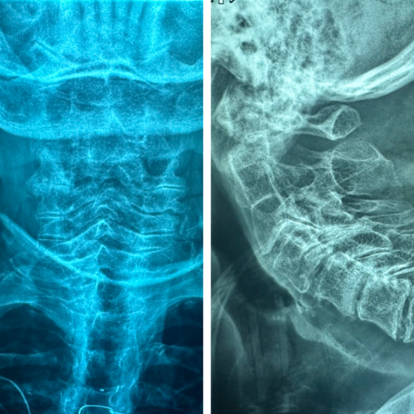
This week in radiology, we saw an 80 year-old man present to urgent care after falling from a standing position at his nursing home while cheering for the Kansas City Chiefs in their latest victory. After the fall, his neck was hurting, and nursing home staff called emergency services worried that he might have broken his neck. The man arrived to the emergency room in a cervical collar. The patient's physician ordered plain films to clear his cervical spine.
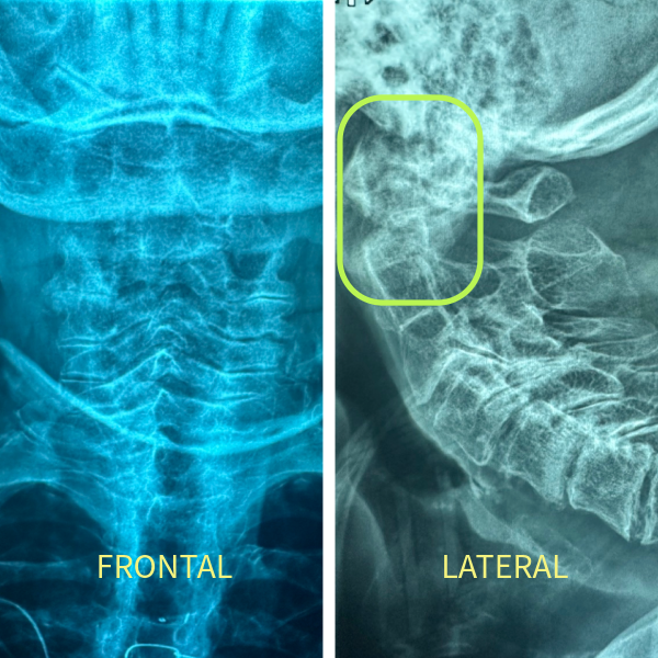
Take a look at the radiographs above. Can you spot the fracture? It's not easy, because the bones overlap in the upper spine. The image on the left is the frontal projection, and the image on the right is the lateral projection. Usually, the frontal projection is taken with the mouth open to move the mandible out of the way. This lets you assess C1-C2 alignment. But this frontal image was taken with a closed mouth, which makes it hard to see what's happening with C1 and C2. The lateral image suggests that something odd is happening with C2.
After reviewing these plain films, the patient's physician ordered a CT c-spine without contrast, which is a good next step when the plain film looks suspicious. It's also reasonable to move to CT directly if your suspicion for injury is high. Similarly, if you ordered a plain film that was normal (aka negative), you would still proceed to CT if your concern for injury was still high, recognizing that plain film can easily hide subtle but important injuries. Check out the CT scan below. Can you see the fracture now?
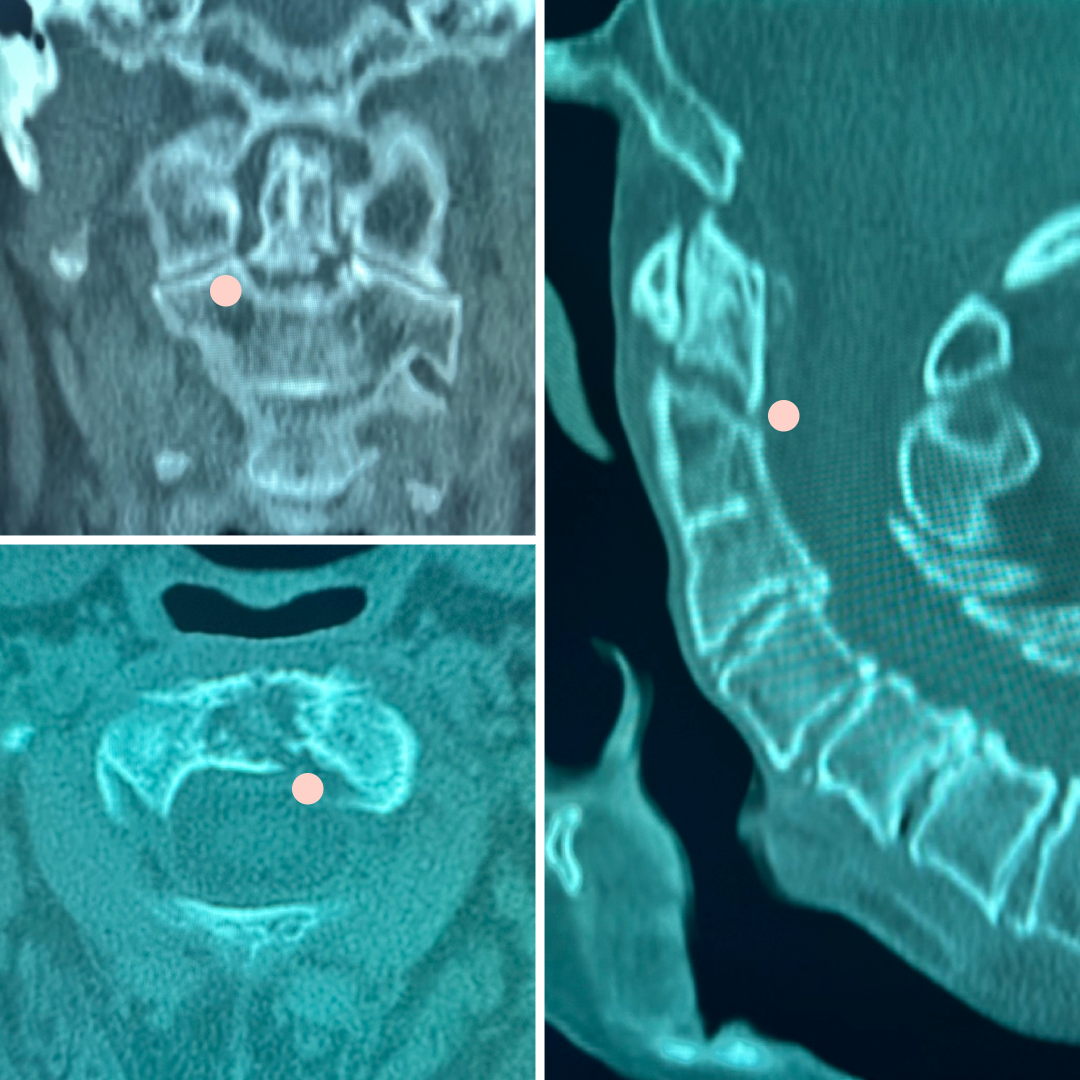
The CT confirms there is a fracture through the upper portion of the C2 vertebral body, called the dens or odontoid process. The dens projects upwards and slides into the C1 vertebral body, allowing your spine the flexibility it needs for you to nod your head back and forth when you're saying "no".
Diagnosing injury to the cervical spine is critical to protect your patient against devastating injury to the cervical spinal cord. Radiographs of the cervical spine are frequently used as a first-line assessment, but c-spine radiographs can hide critical findings. Going straight to a CT cervical spine without contrast is perfectly reasonable when your suspicion is high based on history or physical exam. MR cervical spine without contrast is an important additional study when you're considering ligamentous injury. With experience, you'll learn to select these imaging alternatives in concert with your clinical assessment.
—RadClerk Team
Our cases are provided for educational purposes. Elements of the patient's history have been modified for privacy while retaining a realistic clinical presentation relative to the radiological findings. Any decision-making for specific patients on your service should be made by the patient's expert team, accounting for the specific context of that patient and practice environment.