The case of holiday decorations
This week in radiology, we saw a woman present to urgent care with foot pain after falling from a ladder while hanging lights for the holidays.
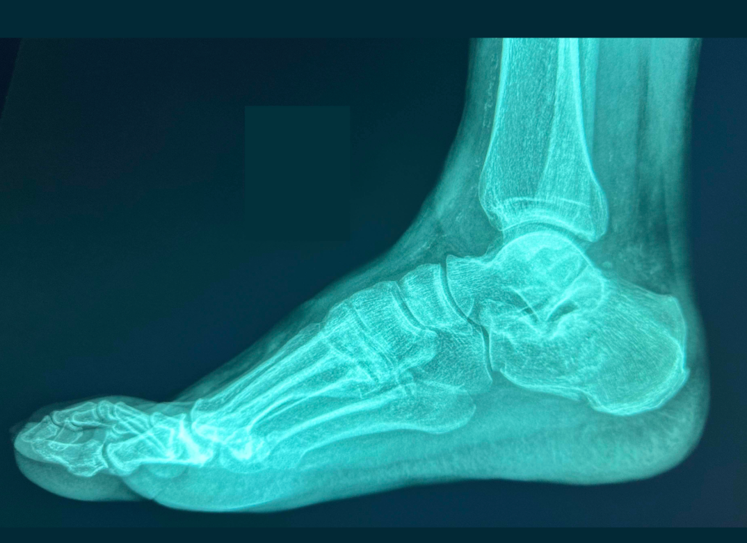
This week in radiology, we saw a woman present to urgent care with foot pain after falling from a ladder while hanging lights for the holidays. After the fall, she was unable to bear weight on her right foot, and came in to urgent care for diagnosis and treatment. In this case, a three-view foot radiograph is a good way to detect fractures, but the foot radiograph is a landmine for missed fractures.
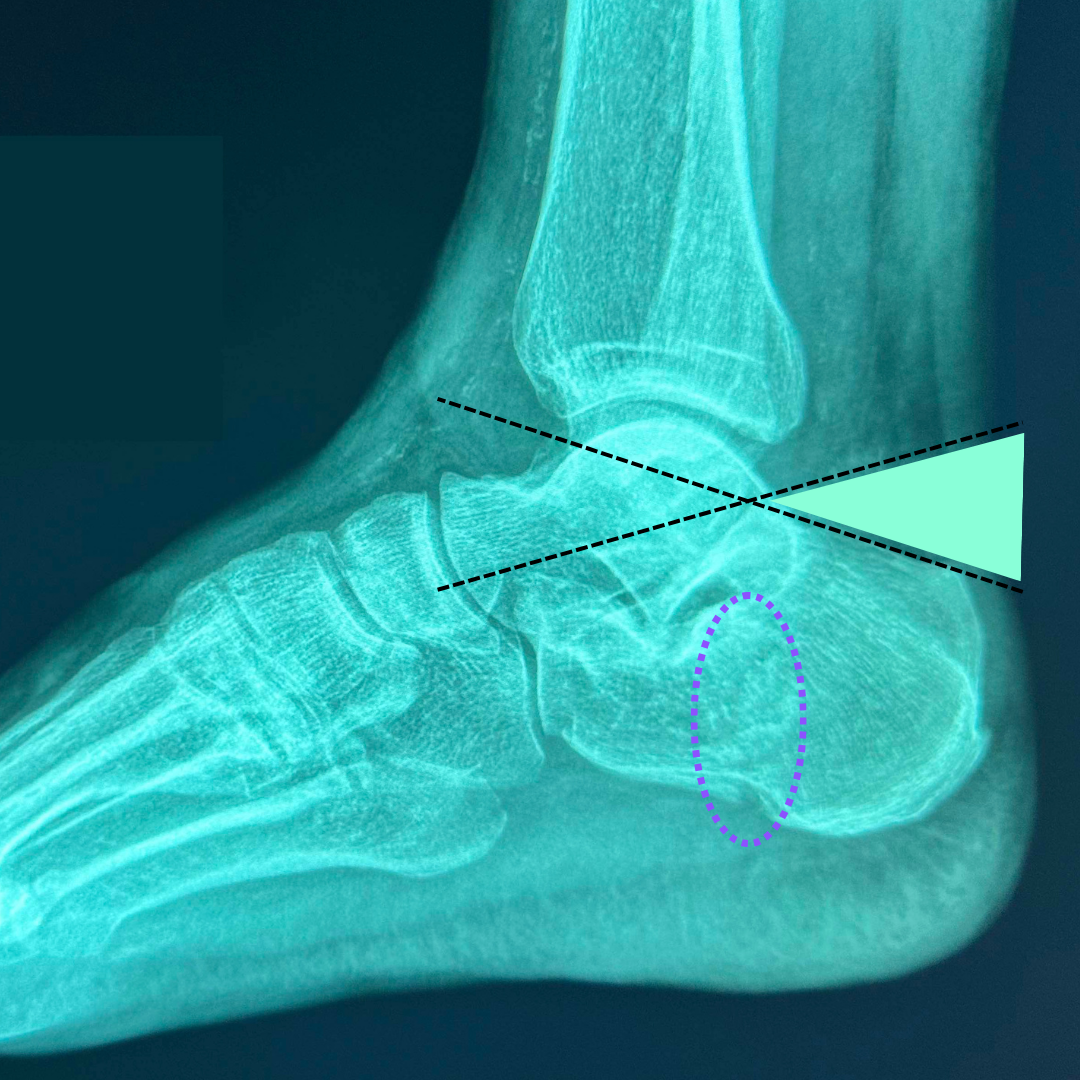
Take a look at the lateral projection above. Can you spot the fracture? This is a nondisplaced calcaneal fracture, which is one of the harder fractures to detect. In this case, we have two clues that there's a calcaneal fracture, which are marked on the annotated image below.
- Subtle cortical disruption. There's a subtle break in the cortical bone at the plantar aspect of the calcaneus (dotted purple circle).
- Flattened calcaneus. Second, the dorsal surface of the calcaneus looks flattened. The black dotted lines follow the posterior and anterior portions of the superior margin of the calcaneus. The angle between them (green triangle) is the Böhler angle, named after Austrian surgeon Lorenz Böhler (1885-1973). When the Böhler angle is less than 25°, you should suspect a calcaneal fracture.
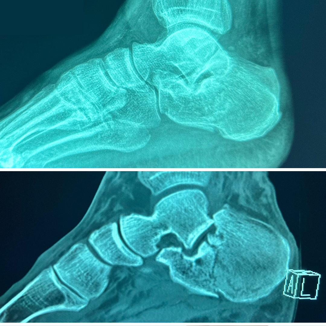
Comparing across modalities is a great way to improve your skills with projection radiography. Below, you can see the radiograph right next to a sagittal reconstruction through the foot on CT. The CT confirms the fracture and helps more thoroughly evaluate the foot for other subtle fractures.
Ordering a radiograph after trauma to the foot is a good first-line assessment. The foot radiograph is one of the more challenging studies for detecting fractures, but with practice, you'll learn to see the different fracture patterns that can hide in plain sight.
—RadClerk Team